Surgical management traditionally involves a Z-plasty or V-to-Y plasty of the medial canthus to increase the length of the vertical scar thus. 1 We report a.

Medial Canthal Webbing Seen After Upper Lid Blepharoplasy Done By A Download Scientific Diagram
Canthal web revision Canthoplasty Revision Canthoplasty The area where the upper and lower lids meet is called the canthus.
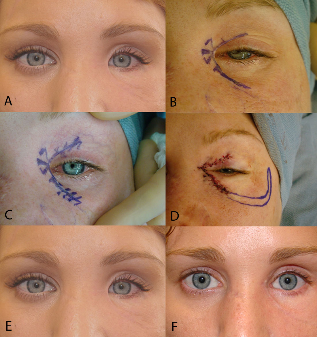
. All clinical photographs are actual patients of Dr. And blepharoplasty represent the commonest iatrogenic causes of medial canthal webbing. I recommend Z-plasty repair of the medial canthal webbing.
Canthal tilt was considered positive when the ies have analyzed the complication rates of lower lateral canthus was superior to the level of medial blepharoplasty after routine canthal support45 canthus neutral when it was located at the same Since 1994 the senior author MAC has per- level and negative when it was inferior Fig. Seven 88 of the patients had lateral canthal webs after surgery and 1 12 patient had a medial canthal web after a motor vehicle accident. The skin then bridges the superomedial hollow of the upper lid in a straight line.
This area near the nose is called the medial canthus and the same area on the outer eyelids is called the lateral canthus. Medial canthus Mohs defect. Epicanthal webbing can result from both traumatic and iatrogenic injuries.
Canthal webs occur because there is a relative deficiency of skin in the vertical as compared to the horizontal plane. Ad When it comes to blepharoplasty or surgery of the eyelids there are certain things that. An effective preventive measure is to taper the nasal incision markings superiorly and to avoid extending the marking medially to the punctum.
Canthal webbing can be associated with scleral show laterally due to inferior lid retraction and is a known complication of blepharoplasty or reconstruction following trauma or tumour excision. Medial skin redundancy when present is best managed by angling the skin marking superiorly 3 to 4 mm medial to the superior punctum. Early recognition and aggressive massage will eliminate the majority of cases.
Anatomy of the upper eyelid. After glabellar flap repair. The latter is usually related to skin incisions required to gain external access to the ethmoidal and frontal sinuses or more rarely to the lacrimal sac.
They argue that canthal anchoring sutures are not needed permanently and using absorbable sutures prevents the risk of stitch sinus or infection. If the marking is angled inferiorly aesthetically displeasing medial canthal webbing may occur and the incision is more noticeable. Medial canthus Mohs defect.
Download high-res image 98KB Download. Medial canthal web after upper blepharoplasty. Large basal cell carcinoma.
Persistent cases are treated by a V- to-Y plasty procedure. Most plastic surgeons avoid addressing canthal webs as they are difficult to. Webs abnormal folds of skin can occur in both areas and are referred to as medial and lateral canthal webs.
Aggressive excision of medial eyelid skin including some nasal skin spectacle deformation of the medial canthus and a preexisting tarsal fold increase the risk of web formation. The rhomboid flap is an effective quick and simple technique for medial canthal reconstruction. Every patient voiced aesthetic concerns with the web and 4 57 of the 7 patients with lateral canthal webs.
There were no major complications or re-operations. May be due to inadvertent trauma to the levator complex including postsurgical edema and dehiscence. The scars usually occur when the incisions are carried too medially and the skin bridges the supero-medial hollow of the upper lid in a straight line.
Less Sorry about your problem. Everyone has seen their eyes be tired and puffy but some people have to live with that. The skin and orbicularis oculi muscle form the anterior layers of the upper eyelid.
It provides excellent cosmesis and is associated with minimal complications. The cosmetic result was highly satisfactory in all cases. A vertical lengthening procedure can correct the lateral canthal web.
Complications such as lagophthalmos lid retraction hematoma injury to extraocular muscles and retrobulbar hemorrhage can occur following blepharoplasty. Abstract Secondary lateral canthal webs may occur after trauma or blepharoplasty. However these complications are uncommon.
Post-blepahroplasty webbing can be seen when upper and lower blepharoplasty is performed together and the lateral skin incisions of the procedures are in close proximity less than 5 mm apart. May be due to incision extended too far medially. Canthal webbing occurs when incisions are car- ried too medially as seen in Figure 9.
WARNING Some photos may be explicit. If it persists then revision by Y-Vtype incisions is warranted. Postoperative canthal webbing is aesthetically displeasing and may be noticed by the patient andor surgeon.
December 11 2018 Answer. Massage and steroid injections can help. Medial canthal webbing Attention to a few important surgical guidelines regarding medial upper eyelid wound closure will help prevent webbing in the medial canthus.
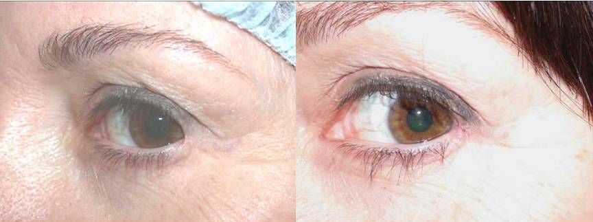
If a medial canthal web does result time massage and steroid injections can help. The best way to handle it is to do a push-pull massage of the surgical area. Medial canthal webbing can be revised with a Z-plasty.
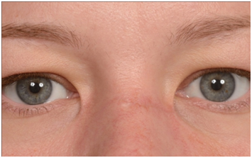
Although it appears that you have subtle webbing this appearance may be quite different after complete healing. They are surgically corrected by a variety of micro-skin flaps. Two cases had minor webbing of the medial upper lid.
Suture is passed through the lateral canthal tendon and suspended superiorly up through an upper eyelid blepharoplasty incision. May be corrected by Zplasty Wplasty transposition flaps or YV advancement procedures. Webbing 3 weeks after blepharoplasty.
Do it at least 3 times a day with 3 repeatitions and 20-30 seconds hold in each more Sorry about your problem. The skin then bridges the superomedial hollow of the upper lid in a straight line. Prevent by planning an incision that extends to the medial commissure.
In cases of canthoplasty the upper and lower eyelid should be carefully and precisely aligned with 6-0 buried vicryl sutures to prevent canthal webbing. A thorough understanding of the upper eyelid anatomy is essential when evaluating patients for possible upper blepharoplasty. A lateral canthal web may form as a consequence of vertical tissue insufficiency after blepharoplasty.
In some cases early recognition and aggressive massage can result in better aesthetic results by improving. After glabellar flap repair. Periosteal flap Leone Am J Ophthalmol 1992 After exposure of the lateral orbital rim a tongue of periosteum is incised in.
At this point after surgery it is normal to have thickening and redness of the incision line. Medial canthal webbing occurs when incisions are carried too medially as seen in Figure 9. This quite common complication following a blepharoplasty operation can be triggered by several factors such as intraoperative canthopexy postoperative temporary lagophthalmos concurrent upper and lower blepharoplasty and transcutaneous approaches violating the orbicularis oculi muscle.
Large basal cell carcinoma. Deep to these layers is the orbital septum which originates from the arcus marginalis at the superior.
Case Of Bilateral Iatrogenic Medial Canthal Webbing Treated With Full Thickness Skin Grafts Medcrave Online

Medial Canthal Webbing Seen After Upper Lid Blepharoplasy Done By A Download Scientific Diagram

Medial Canthal Webbing Seen After Upper Lid Blepharoplasy Done By A Download Scientific Diagram

Medial Canthal Webbing Seen After Upper Lid Blepharoplasy Done By A Download Scientific Diagram
Canthal Web Revision Canthoplasty Revision Canthoplasty Dr Guy Massry
![]()
Medial Canthal Webbing Seen After Upper Lid Blepharoplasy Done By A Download Scientific Diagram
Case Of Bilateral Iatrogenic Medial Canthal Webbing Treated With Full Thickness Skin Grafts Medcrave Online
Revisional Eyelid Surgery Fixing The Canthus Dr Guy Massry
0 comments
Post a Comment